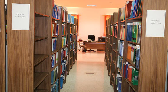
BibliothĆØque de la FMPO
 . Bibliothèque
 Radiologie

 Affiner la recherche
Savoir Faire en Radiologie OstƩo-Articulaire Num 11 : Index Bibliographique de la Collection / Jean-Denis Laredo
Titre : Savoir Faire en Radiologie OstĆ©o-Articulaire Num 11 : Index Bibliographique de la Collection Type de document : texte imprimĆ© Auteurs : Jean-Denis Laredo, Auteur ; M. Wybier, Auteur ; Collectif, Auteur Editeur : Sauramps MĆ©dical AnnĆ©e de publication : 2009 ISBN/ISSN/EAN : 978-2-84023-616-0 Langues : FranƧais (fre) CatĆ©gories : Radiologie RĆ©sumĆ© : Cet ouvrage est le onziĆØme numĆ©ro (2009) de la collection SAVOIR-FAIRE en radiologie ostĆ©o-articulaire. Le principe du Savoir-Faire en Radiologie OstĆ©o-articulaire est d'apporter une connaissance qui soit directement et rapidement utilisable dans la pratique radiologique quotidienne. Les affections rares sont volontiers laissĆ©es de cĆ´tĆ©, pour traiter la pathologie frĆ©quente dont tous les modes de prĆ©sentations ne sont pas toujours connus. Les sujets traitĆ©s sont dans le champ de la rhumatologie, de l'orthopĆ©die et de la technique radiologique. L'information essaie de couvrir l'ensemble des donnĆ©es utiles, de l'anatomopathologie Ć la clinique, ainsi que, bien sĆ»r, Ć l'imagerie et finalement Ć la thĆ©rapeutique. Nos collĆØgues rhumatologues et chirurgiens orthopĆ©distes participent largement Ć ces enseignements. L'objectif de cette Collection est que le lecteur puisse s'y rapporter quand il sera confrontĆ© Ć un cas pratique, par l'intermĆ©diaire de l'index cumulatif qui permet de retrouver tous les sujets traitĆ©s depuis le premier numĆ©ro (2000). Ce numĆ©ro 11 traite de sujets d'actualitĆ© en imagerie des articulations (Ć©paule, coude, genou, cheville), en pathologie pseudo-tumorale des parties molles, en podologie. Il fait un recensement des complications liĆ©es aux suspensions cortisoniques injectĆ©es, notamment au rachis lombaire. Les avancĆ©es techniques (sĆ©quences d'IRM, intĆ©rĆŖt et complications des chĆ©lates de gadolinium) et les nouvelles dispositions rĆ©glementaires pour la radiologie font aussi partie du programme de cet ouvrage. Savoir Faire en Radiologie OstĆ©o-Articulaire Num 11 : Index Bibliographique de la Collection [texte imprimĆ©] / Jean-Denis Laredo, Auteur ; M. Wybier, Auteur ; Collectif, Auteur . - [S.l.]Ā : Sauramps MĆ©dical, 2009.
ISBN : 978-2-84023-616-0
Langues : FranƧais (fre)
CatĆ©gories : Radiologie RĆ©sumĆ© : Cet ouvrage est le onziĆØme numĆ©ro (2009) de la collection SAVOIR-FAIRE en radiologie ostĆ©o-articulaire. Le principe du Savoir-Faire en Radiologie OstĆ©o-articulaire est d'apporter une connaissance qui soit directement et rapidement utilisable dans la pratique radiologique quotidienne. Les affections rares sont volontiers laissĆ©es de cĆ´tĆ©, pour traiter la pathologie frĆ©quente dont tous les modes de prĆ©sentations ne sont pas toujours connus. Les sujets traitĆ©s sont dans le champ de la rhumatologie, de l'orthopĆ©die et de la technique radiologique. L'information essaie de couvrir l'ensemble des donnĆ©es utiles, de l'anatomopathologie Ć la clinique, ainsi que, bien sĆ»r, Ć l'imagerie et finalement Ć la thĆ©rapeutique. Nos collĆØgues rhumatologues et chirurgiens orthopĆ©distes participent largement Ć ces enseignements. L'objectif de cette Collection est que le lecteur puisse s'y rapporter quand il sera confrontĆ© Ć un cas pratique, par l'intermĆ©diaire de l'index cumulatif qui permet de retrouver tous les sujets traitĆ©s depuis le premier numĆ©ro (2000). Ce numĆ©ro 11 traite de sujets d'actualitĆ© en imagerie des articulations (Ć©paule, coude, genou, cheville), en pathologie pseudo-tumorale des parties molles, en podologie. Il fait un recensement des complications liĆ©es aux suspensions cortisoniques injectĆ©es, notamment au rachis lombaire. Les avancĆ©es techniques (sĆ©quences d'IRM, intĆ©rĆŖt et complications des chĆ©lates de gadolinium) et les nouvelles dispositions rĆ©glementaires pour la radiologie font aussi partie du programme de cet ouvrage. Exemplaires (5)
Code-barres Cote Support Localisation Section DisponibilitĆ© 1503 Radio 080 Livre Bibliothèque Radiologie Disponible 1731 Radio 080 Livre Bibliothèque Radiologie Disponible 1732 Radio 080 Livre Bibliothèque Radiologie Disponible 1733 Radio 080 Livre Bibliothèque Radiologie Disponible 1972 Radio 080 Livre Bibliothèque Radiologie Disponible
Titre : Savoir Faire en Radiologie OstĆ©o-Articulaire Num 5 : Avec Index Cumulatif des Tomes 1 Ć 5 (Genou - PĆ©diatrie - Epaule - Hanche - Tumeurs et Pseudotumeurs - Technique) Type de document : texte imprimĆ© Auteurs : Jean-Denis Laredo, Auteur ; Marc Wybier, Auteur ; Laurence BellaĆÆche, Auteur Editeur : Sauramps MĆ©dical AnnĆ©e de publication : 2003 ISBN/ISSN/EAN : 978-2-84023-056-4 Langues : FranƧais (fre) CatĆ©gories : Radiologie RĆ©sumĆ© : Le principe du savoir faire en radiologie ostĆ©o-articulaire est d'apporter une connaissance qui soit directement et rapidement utilisable dans la pratique radiologique quotidienne. Les affections rares sont volontiers laissĆ©es de cĆ´tĆ© pour traiter la pathologie frĆ©quente dont tous les aspects et tous les modes de prĆ©sentations ne sont pas toujours connus. Les sujets traitĆ©s sont dans le champ de la rhumatologie, de l'orthopĆ©die et de la technique radiologique. L'information essaie de couvrir l'ensemble des donnĆ©es utiles, de l'anatomopathologie Ć la clinique, Ć l'imagerie bien sĆ»r et finalement Ć la thĆ©rapeutique. Nos collĆØgues rhumatologues et chirurgiens orthopĆ©distes participent largement Ć ces enseignements. L'objectif est de faire une collection Ć laquelle le lecteur pourra se reporter quand il sera confrontĆ© Ć un cas pratique en utilisant l'index cumulatif qui permet de retrouver tous les sujets traitĆ©s depuis le premier tome (2000). Ce volume concerne l'imagerie des prothĆØses du genou, l'imagerie de l'Ć©paule (coiffe des rotateurs, bourrelet glĆ©noĆÆdien), de la hanche (interligne articulaire normal et pathologique), des tendons et des muscles (fessiers, ischio-jambiers), des traumatismes (fracture de l'extrĆ©mitĆ© supĆ©rieure de l'humĆ©rus, traumatismes des membres chez l'enfant), du rhumatisme (coxite de l'adulte), de tumeurs et pseudotumeurs (myosite ossifiante circonscrite, lacunes des doigts, lacunes du crĆ¢ne, hĆ©mangiomes des parties molles), et des sujets techniques (densitomĆ©trie osseuse, IRM de la moelle osseuse, Ć©chodoppler des artĆØres, ponction des calcifications de l'Ć©paule).
Savoir Faire en Radiologie OstĆ©o-Articulaire Num 5 : Avec Index Cumulatif des Tomes 1 Ć 5 (Genou - PĆ©diatrie - Epaule - Hanche - Tumeurs et Pseudotumeurs - Technique) [texte imprimĆ©] / Jean-Denis Laredo, Auteur ; Marc Wybier, Auteur ; Laurence BellaĆÆche, Auteur . - [S.l.]Ā : Sauramps MĆ©dical, 2003.
ISBN : 978-2-84023-056-4
Langues : FranƧais (fre)
CatĆ©gories : Radiologie RĆ©sumĆ© : Le principe du savoir faire en radiologie ostĆ©o-articulaire est d'apporter une connaissance qui soit directement et rapidement utilisable dans la pratique radiologique quotidienne. Les affections rares sont volontiers laissĆ©es de cĆ´tĆ© pour traiter la pathologie frĆ©quente dont tous les aspects et tous les modes de prĆ©sentations ne sont pas toujours connus. Les sujets traitĆ©s sont dans le champ de la rhumatologie, de l'orthopĆ©die et de la technique radiologique. L'information essaie de couvrir l'ensemble des donnĆ©es utiles, de l'anatomopathologie Ć la clinique, Ć l'imagerie bien sĆ»r et finalement Ć la thĆ©rapeutique. Nos collĆØgues rhumatologues et chirurgiens orthopĆ©distes participent largement Ć ces enseignements. L'objectif est de faire une collection Ć laquelle le lecteur pourra se reporter quand il sera confrontĆ© Ć un cas pratique en utilisant l'index cumulatif qui permet de retrouver tous les sujets traitĆ©s depuis le premier tome (2000). Ce volume concerne l'imagerie des prothĆØses du genou, l'imagerie de l'Ć©paule (coiffe des rotateurs, bourrelet glĆ©noĆÆdien), de la hanche (interligne articulaire normal et pathologique), des tendons et des muscles (fessiers, ischio-jambiers), des traumatismes (fracture de l'extrĆ©mitĆ© supĆ©rieure de l'humĆ©rus, traumatismes des membres chez l'enfant), du rhumatisme (coxite de l'adulte), de tumeurs et pseudotumeurs (myosite ossifiante circonscrite, lacunes des doigts, lacunes du crĆ¢ne, hĆ©mangiomes des parties molles), et des sujets techniques (densitomĆ©trie osseuse, IRM de la moelle osseuse, Ć©chodoppler des artĆØres, ponction des calcifications de l'Ć©paule).
Exemplaires (2)
Code-barres Cote Support Localisation Section DisponibilitĆ© 885 Radio 078 Livre Bibliothèque Radiologie Disponible 886 Radio 078 Livre Bibliothèque Radiologie Disponible
Titre : "Scanner et IRM Cardiaques PƩdiatriques ""Imagerie MƩdicale Pratique"" : Cardiopathies CongƩnitales - Descriptions Anatomocliniques - Protocoles d'Exploration - Analyses Radiologique " Type de document : texte imprimƩ Auteurs : Phalla OU, Auteur ; Daniel Sidi, Auteur ; Damien Bonnet, Auteur Editeur : Elsevier Masson AnnƩe de publication : 2008 ISBN/ISSN/EAN : 978-2-294-07465-3 Langues : FranƧais (fre) CatƩgories : Radiologie RƩsumƩ : Les techniques d'imagerie en coupes - scanner multidƩtecteurs et IRM - sont aujourd'hui des mƩthodes d'exploration indispensables pour une Ʃvaluation moderne des cardiopathies congƩnitales. Fruit d'une collaboration Ʃtroite entre radiologues, cardiologues et chirurgiens cardiovasculaires, cet ouvrage fait le point sur l'apport de l'imagerie en coupe moderne en cardiologie pƩdiatrique. L'ouvrage fait un rappel des principes techniques et expose les spƩcificitƩs de chacune des mƩthodes d'exploration.
Les principales anomalies congĆ©nitales sont prĆ©sentĆ©es de faƧon brĆØve et claire: anomalies des artĆØres coronaires, de l'aorte, des artĆØres pulmonaires et des retours veineux, pathologies du myocarde, tumeurs cardiaques primitives, etc. Les indications radiologiques et les protocoles d'examen sont dĆ©taillĆ©s de maniĆØre pratique. L'abondance des illustrations aidera le praticien dans sa dĆ©marche diagnostique: plus de 400 clichĆ©s, graphiques et schĆ©mas ont Ć©tĆ© sĆ©lectionnĆ©s pour leur aspect didactique.
Cet ouvrage constitue un outil essentiel pour les radiologues, cardiologues et chirurgiens cardiovasculaires impliquĆ©s dans la prise en charge des cardiopathies congĆ©nitales. Il intĆ©ressera Ć©galement les pĆ©diatres et nĆ©onatologistes-rĆ©animateurs. En outre, les fiches pratiques sur les techniques d'examen offrent un support prĆ©cieux pour les manipulateurs."Scanner et IRM Cardiaques PĆ©diatriques ""Imagerie MĆ©dicale Pratique"" : Cardiopathies CongĆ©nitales - Descriptions Anatomocliniques - Protocoles d'Exploration - Analyses Radiologique " [texte imprimĆ©] / Phalla OU, Auteur ; Daniel Sidi, Auteur ; Damien Bonnet, Auteur . - [S.l.]Ā : Elsevier Masson, 2008.
ISBN : 978-2-294-07465-3
Langues : FranƧais (fre)
CatƩgories : Radiologie RƩsumƩ : Les techniques d'imagerie en coupes - scanner multidƩtecteurs et IRM - sont aujourd'hui des mƩthodes d'exploration indispensables pour une Ʃvaluation moderne des cardiopathies congƩnitales. Fruit d'une collaboration Ʃtroite entre radiologues, cardiologues et chirurgiens cardiovasculaires, cet ouvrage fait le point sur l'apport de l'imagerie en coupe moderne en cardiologie pƩdiatrique. L'ouvrage fait un rappel des principes techniques et expose les spƩcificitƩs de chacune des mƩthodes d'exploration.
Les principales anomalies congĆ©nitales sont prĆ©sentĆ©es de faƧon brĆØve et claire: anomalies des artĆØres coronaires, de l'aorte, des artĆØres pulmonaires et des retours veineux, pathologies du myocarde, tumeurs cardiaques primitives, etc. Les indications radiologiques et les protocoles d'examen sont dĆ©taillĆ©s de maniĆØre pratique. L'abondance des illustrations aidera le praticien dans sa dĆ©marche diagnostique: plus de 400 clichĆ©s, graphiques et schĆ©mas ont Ć©tĆ© sĆ©lectionnĆ©s pour leur aspect didactique.
Cet ouvrage constitue un outil essentiel pour les radiologues, cardiologues et chirurgiens cardiovasculaires impliquƩs dans la prise en charge des cardiopathies congƩnitales. Il intƩressera Ʃgalement les pƩdiatres et nƩonatologistes-rƩanimateurs. En outre, les fiches pratiques sur les techniques d'examen offrent un support prƩcieux pour les manipulateurs.Exemplaires (1)
Code-barres Cote Support Localisation Section DisponibilitĆ© 6275 Radio 225 Livre Bibliothèque Radiologie Disponible
Titre : Skeletal Development of the Hand and Wrist : A Radiographic Atlas & digital Bone Age Companion Type de document : texte imprimƩ Auteurs : Cree M. Gaskin, Auteur ; S. Lowell Kahn, Auteur Editeur : Oxford AnnƩe de publication : 2011 ISBN/ISSN/EAN : 978-0-19-978205-5 Langues : FranƧais (fre) CatƩgories : Radiologie RƩsumƩ : Bone age assessment, a crucial part of the diagnosis and management of pediatric growth disorders as well as the timing of certain pediatric orthopedic procedures, has for decades depended on the meticulous examination of plain radiographs. Examining the subtle changes present within the maturing human hand often proves to be challenging and time-consuming.
Building on the popular Greulich and Pyle atlas, this book modernizes the method for pediatric skeletal maturity determination. It offers a wealth of images, carefully mined from thousands of digital radiographs from University of Virginia's Picture Archiving and Communication System (PACS), edited to best demonstrate important developmental bone features, and organized by age and sex for rapid reference. To expedite learning and clinical image analysis, images come in pairs: annotated and unannotated, for easy comparison. Succinct annotations on the images replace lengthy text to provide a quicker and clearer understanding of the skeletal age. These annotations highlight important and subtle features to help distinguish images that otherwise look superficially alike. The result is an atlas of exceptionally high quality skeletal radiographic standards that capture both the major and finer details of the accepted standards of Greulich and Pyle.
The user-friendly format of this book enables a faster, more accurate, and more educational approach to determining skeletal maturity. The Digital Bone Age Companion packaged with the book is a computer program that facilitates viewing of the atlas images in digital format. Users can easily zoom in on radiographic features, set image level and width to their preference, and compare two or three reference standards side-by-side for difficult cases. Most importantly, the program expedites evaluation, optimizes workflow, and minimizes user-introduced errors with the reliable bone age calculator and built-in report generator. The digital format may also be available for integration with your Radiology Information System (RIS) for further workflow enhancement.
Given the broad application of pediatric bone aging, Skeletal Development of the Hand and Wrist is not only intended for practicing and training radiologists, but for all of those who employ bone age studies as part of their practice.Skeletal Development of the Hand and Wrist : A Radiographic Atlas & digital Bone Age Companion [texte imprimĆ©] / Cree M. Gaskin, Auteur ; S. Lowell Kahn, Auteur . - [S.l.]Ā : Oxford, 2011.
ISBN : 978-0-19-978205-5
Langues : FranƧais (fre)
CatƩgories : Radiologie RƩsumƩ : Bone age assessment, a crucial part of the diagnosis and management of pediatric growth disorders as well as the timing of certain pediatric orthopedic procedures, has for decades depended on the meticulous examination of plain radiographs. Examining the subtle changes present within the maturing human hand often proves to be challenging and time-consuming.
Building on the popular Greulich and Pyle atlas, this book modernizes the method for pediatric skeletal maturity determination. It offers a wealth of images, carefully mined from thousands of digital radiographs from University of Virginia's Picture Archiving and Communication System (PACS), edited to best demonstrate important developmental bone features, and organized by age and sex for rapid reference. To expedite learning and clinical image analysis, images come in pairs: annotated and unannotated, for easy comparison. Succinct annotations on the images replace lengthy text to provide a quicker and clearer understanding of the skeletal age. These annotations highlight important and subtle features to help distinguish images that otherwise look superficially alike. The result is an atlas of exceptionally high quality skeletal radiographic standards that capture both the major and finer details of the accepted standards of Greulich and Pyle.
The user-friendly format of this book enables a faster, more accurate, and more educational approach to determining skeletal maturity. The Digital Bone Age Companion packaged with the book is a computer program that facilitates viewing of the atlas images in digital format. Users can easily zoom in on radiographic features, set image level and width to their preference, and compare two or three reference standards side-by-side for difficult cases. Most importantly, the program expedites evaluation, optimizes workflow, and minimizes user-introduced errors with the reliable bone age calculator and built-in report generator. The digital format may also be available for integration with your Radiology Information System (RIS) for further workflow enhancement.
Given the broad application of pediatric bone aging, Skeletal Development of the Hand and Wrist is not only intended for practicing and training radiologists, but for all of those who employ bone age studies as part of their practice.Exemplaires (1)
Code-barres Cote Support Localisation Section DisponibilitĆ© 5112 Radio 197 Livre Bibliothèque Radiologie Disponible
Titre : Le Spermocytogramme en Images Type de document : texte imprimƩ Auteurs : Alexandra Mesner, Auteur ; Catherine Poirot, Auteur ; Pierre Jouannet, Auteur Editeur : Editions Vernazobres-Grego AnnƩe de publication : 2010 ISBN/ISSN/EAN : 978-2-84136-889-1 Langues : FranƧais (fre) CatƩgories : Biologie
GynƩcologie
RadiologieRĆ©sumĆ© : FormĆ© dans le testicule suite Ć un ensemble de divisions et de diffĆ©renciations cellulaires et de phĆ©nomĆØnes molĆ©culaires aussi complexes que fragiles, le spermatozoĆÆde doit quitter l'organe et l'ĆŖtre qui sont Ć son origine pour aller cheminer chez un autre Ć la rencontre de sa partenaire fĆ©minine, l'ovocyte, afin d'initier la formation d'un 3e ĆŖtre, l'enfant. Destin bien particulier qui amĆØne les spermatozoĆÆdes Ć se confronter Ć des espaces, des milieux, des interactions les plus divers et quelquefois mĆŖme les plus hostiles. Destin bien particulier car pratiquement aucun d'entre eux ne sera en mesure d'accomplir l'Ć©tape ultime de la mission pour laquelle il a Ć©tĆ© conƧu. Pourtant tout, en principe, a Ć©tĆ© fait pour y arriver. En effet, le spermatozoĆÆde rĆ©sulte d'une diffĆ©renciation cellulaire la plus extrĆŖme au seul service de sa finalitĆ© : la fĆ©condation. Le gĆ©nome haploĆÆde et unique qui a Ć©tĆ© fabriquĆ© grĆ¢ce Ć la mĆ©iose a Ć©tĆ© emballĆ© dans des protĆ©ines spĆ©cifiques qui ont pour mission de le protĆ©ger mais aussi d'empĆŖcher l'expression intempestive de ses gĆØnes. Puis il a fallu construire des systĆØmes de transport, de reconnaissance et de fusion trĆØs sophistiquĆ©s pour interagir avec l'ovocyte et seulement avec lui. La construction cellulaire et molĆ©culaire du flagelle, de l'acrosome et de la membrane qui permet d'atteindre cet objectif correspond Ć la phase ultime de la spermatogenĆØse : la spermiogenĆØse.
Le Spermocytogramme en Images [texte imprimĆ©] / Alexandra Mesner, Auteur ; Catherine Poirot, Auteur ; Pierre Jouannet, Auteur . - [S.l.]Ā : Editions Vernazobres-Grego, 2010.
ISBN : 978-2-84136-889-1
Langues : FranƧais (fre)
CatƩgories : Biologie
GynƩcologie
RadiologieRĆ©sumĆ© : FormĆ© dans le testicule suite Ć un ensemble de divisions et de diffĆ©renciations cellulaires et de phĆ©nomĆØnes molĆ©culaires aussi complexes que fragiles, le spermatozoĆÆde doit quitter l'organe et l'ĆŖtre qui sont Ć son origine pour aller cheminer chez un autre Ć la rencontre de sa partenaire fĆ©minine, l'ovocyte, afin d'initier la formation d'un 3e ĆŖtre, l'enfant. Destin bien particulier qui amĆØne les spermatozoĆÆdes Ć se confronter Ć des espaces, des milieux, des interactions les plus divers et quelquefois mĆŖme les plus hostiles. Destin bien particulier car pratiquement aucun d'entre eux ne sera en mesure d'accomplir l'Ć©tape ultime de la mission pour laquelle il a Ć©tĆ© conƧu. Pourtant tout, en principe, a Ć©tĆ© fait pour y arriver. En effet, le spermatozoĆÆde rĆ©sulte d'une diffĆ©renciation cellulaire la plus extrĆŖme au seul service de sa finalitĆ© : la fĆ©condation. Le gĆ©nome haploĆÆde et unique qui a Ć©tĆ© fabriquĆ© grĆ¢ce Ć la mĆ©iose a Ć©tĆ© emballĆ© dans des protĆ©ines spĆ©cifiques qui ont pour mission de le protĆ©ger mais aussi d'empĆŖcher l'expression intempestive de ses gĆØnes. Puis il a fallu construire des systĆØmes de transport, de reconnaissance et de fusion trĆØs sophistiquĆ©s pour interagir avec l'ovocyte et seulement avec lui. La construction cellulaire et molĆ©culaire du flagelle, de l'acrosome et de la membrane qui permet d'atteindre cet objectif correspond Ć la phase ultime de la spermatogenĆØse : la spermiogenĆØse.
Exemplaires (4)
Code-barres Cote Support Localisation Section DisponibilitĆ© 6387 Biologie 42 Livre Bibliothèque Biologie Exclu du prĆŖt 4042 Gyneco 071 Livre Bibliothèque Gynécologie Disponible 4043 Gyneco 071 Livre Bibliothèque Gynécologie Disponible 6679 Radio 250 Livre Bibliothèque Radiologie Disponible
Titre : Squire's Fundamentals of Radiology. 6th ED Type de document : texte imprimĆ© Auteurs : Robert A. Novelline, Auteur Editeur : Harvard University Press AnnĆ©e de publication : 2004 ISBN/ISSN/EAN : 978-0-674-01279-0 Langues : FranƧais (fre) CatĆ©gories : Radiologie RĆ©sumĆ© : In the past five years, the development of new imaging technologies that make possible faster and more accurate diagnoses has significantly improved the imaging of disease and injury. This new edition of Squireā€™s Fundamentals of Radiology describes and illustrates these new techniques to prepare medical students and other radiology learners to provide the most optimal and up-to-date imaging management for their patients. Not only are new diagnostic techniques outlined, such as the multidetector computed tomography diagnosis of pulmonary embolism and the diffusion-weighted magnetic-resonance imaging of stroke, but hundreds of new diagnostic images have been included to illustrate the radiological characteristics of common diseases with state-of-the-art computed radiography, ultrasound, multidetector computed tomography, and magnetic-resonance images. The text has been completely reviewed and updated to present the latest and best strategies in diagnostic imaging.
New interventional radiology procedures have been added, including vertebroplasty, a percutaneous injection treatment of painful spinal compression fractures; uterine artery embolization, a surgical alternative to hysterectomy in women with painful or bleeding uterine fibroids; and radiofrequency ablation, a percutaneous technique for treating unresectable tumors in the liver and other organs with probes that superheat and thus destroy cancer cells.
A new chapter on advances in diagnostic imaging describes many cutting-edge imaging technologies, such as three-dimensional and digital imaging, functional magnetic-resonance imaging, PETā€“CT (positron emission tomography combined with computed tomography), cardiac calcium CT scoring, multidetector gated cardiac CT, and molecular imaging.Squire's Fundamentals of Radiology. 6th ED [texte imprimĆ©] / Robert A. Novelline, Auteur . - [S.l.]Ā : Harvard University Press, 2004.
ISBN : 978-0-674-01279-0
Langues : FranƧais (fre)
CatĆ©gories : Radiologie RĆ©sumĆ© : In the past five years, the development of new imaging technologies that make possible faster and more accurate diagnoses has significantly improved the imaging of disease and injury. This new edition of Squireā€™s Fundamentals of Radiology describes and illustrates these new techniques to prepare medical students and other radiology learners to provide the most optimal and up-to-date imaging management for their patients. Not only are new diagnostic techniques outlined, such as the multidetector computed tomography diagnosis of pulmonary embolism and the diffusion-weighted magnetic-resonance imaging of stroke, but hundreds of new diagnostic images have been included to illustrate the radiological characteristics of common diseases with state-of-the-art computed radiography, ultrasound, multidetector computed tomography, and magnetic-resonance images. The text has been completely reviewed and updated to present the latest and best strategies in diagnostic imaging.
New interventional radiology procedures have been added, including vertebroplasty, a percutaneous injection treatment of painful spinal compression fractures; uterine artery embolization, a surgical alternative to hysterectomy in women with painful or bleeding uterine fibroids; and radiofrequency ablation, a percutaneous technique for treating unresectable tumors in the liver and other organs with probes that superheat and thus destroy cancer cells.
A new chapter on advances in diagnostic imaging describes many cutting-edge imaging technologies, such as three-dimensional and digital imaging, functional magnetic-resonance imaging, PETā€“CT (positron emission tomography combined with computed tomography), cardiac calcium CT scoring, multidetector gated cardiac CT, and molecular imaging.Exemplaires (1)
Code-barres Cote Support Localisation Section DisponibilitĆ© 3785 Radio 083 Livre Bibliothèque Radiologie Disponible
Titre : Structure Radiologique de Base de l'OMS. Manuel d'InterprƩtation Radiographique pour GƩnƩralistes Type de document : texte imprimƩ Auteurs : P. E. S. Palmer, Auteur ; W. P. Cockshott, Auteur Editeur : OMS AnnƩe de publication : 1985 ISBN/ISSN/EAN : 978-92-4-254177-9 Langues : FranƧais (fre) CatƩgories : Radiologie RƩsumƩ : Preface
Acknowlegments
Chapter 1. introdction and basic concepts
Chapter 2. An invitation to think three-dimensionally
Chapter 3. How to study a chast film
Chapter 4. The lung itself
Chapter 5. The roentgen signs of lung consolidation-air space disease
Chapter 6. The diaphragm and the pleural space
Chapter 7. Mediastinal shift, atelectasis and emphysema
Chapter 8. Study of the mediastinal structures
Chapter 9. How to gather and use roentgen data
Chapter 10. The heart
Chapter 11. The abdomen: study of the plain film
Chapter 12. The abdomen: systematic examination of soft tissue zones on the plain film
Chapter 13. The abdomen: distended stomach, large and small bowel; free air and free fluid
Chapter 14. Contrast studies: principles, applications and scope
Chapter 15. Special deductions possible from excretory and secretory contrast studies
Chapter 16. Skull and bones
Structure Radiologique de Base de l'OMS. Manuel d'InterprĆ©tation Radiographique pour GĆ©nĆ©ralistes [texte imprimĆ©] / P. E. S. Palmer, Auteur ; W. P. Cockshott, Auteur . - [S.l.]Ā : OMS, 1985.
ISBN : 978-92-4-254177-9
Langues : FranƧais (fre)
CatƩgories : Radiologie RƩsumƩ : Preface
Acknowlegments
Chapter 1. introdction and basic concepts
Chapter 2. An invitation to think three-dimensionally
Chapter 3. How to study a chast film
Chapter 4. The lung itself
Chapter 5. The roentgen signs of lung consolidation-air space disease
Chapter 6. The diaphragm and the pleural space
Chapter 7. Mediastinal shift, atelectasis and emphysema
Chapter 8. Study of the mediastinal structures
Chapter 9. How to gather and use roentgen data
Chapter 10. The heart
Chapter 11. The abdomen: study of the plain film
Chapter 12. The abdomen: systematic examination of soft tissue zones on the plain film
Chapter 13. The abdomen: distended stomach, large and small bowel; free air and free fluid
Chapter 14. Contrast studies: principles, applications and scope
Chapter 15. Special deductions possible from excretory and secretory contrast studies
Chapter 16. Skull and bones
Exemplaires (2)
Code-barres Cote Support Localisation Section DisponibilitĆ© 3801 Radio 119 Livre Bibliothèque Radiologie Disponible 3802 Radio 119 Livre Bibliothèque Radiologie Disponible
Titre : TDM et IRM Cliniques : Indications et SĆ©miologie de la TomodensitomĆ©trie et de l'Imagerie par RĆ©sonance MagnĆ©tique Type de document : texte imprimĆ© Auteurs : D. Buthiau, Auteur Editeur : Frison-Roche AnnĆ©e de publication : 1992 ISBN/ISSN/EAN : 978-2-87671-160-0 Langues : FranƧais (fre) CatĆ©gories : Radiologie RĆ©sumĆ© : La TomodensitomĆ©trie et la RĆ©sonance MagnĆ©tique se placent certainement au premier rang parmi les techniques dā€™exploration diagnostique qui ont le plus bouleversĆ© nos habitudes de raisonnementā€¦
Cependant, pour ĆŖtre performants, ces procĆ©dĆ©s dā€™imagerie doivent ĆŖtre utilisĆ©s Ć bon escient, les indications correctement posĆ©es, la technique irrĆ©prochable et lā€™interprĆ©tation intelligenteā€¦
A notre Ć©poque ou le temps disponible est le bien le plus rare et le plus cher, il faut que chacun Ć©tablisse une hiĆ©rarchie dans les urgences et les besoins et une vĆ©ritable comptabilitĆ© de ses investissements intellectuelsā€¦
A ce titre, nous pensons que cet ouvrage possĆØde la qualitĆ© dā€™ĆŖtre utile au spĆ©cialiste dā€™organe ou dā€™appareil, au radiologue, au gĆ©nĆ©raliste ou Ć lā€™internisteā€¦
La rĆ©ponse ou une aide Ć la rĆ©flexion ainsi que lā€™interprĆ©tation radiologique trĆØs complĆØte et Ć©laborĆ©e pourrait ĆŖtre fournies par la lecture de ce livre dont la clartĆ© dā€™exposition est une qualitĆ© majeureā€¦
Ce livre sera, nous les prĆ©sumons, dā€™une utilisation quasi quotidienne et constituera un vĆ©ritable outil de travail et de perfectionnement.
TDM et IRM Cliniques : Indications et SĆ©miologie de la TomodensitomĆ©trie et de l'Imagerie par RĆ©sonance MagnĆ©tique [texte imprimĆ©] / D. Buthiau, Auteur . - [S.l.]Ā : Frison-Roche, 1992.
ISBN : 978-2-87671-160-0
Langues : FranƧais (fre)
CatĆ©gories : Radiologie RĆ©sumĆ© : La TomodensitomĆ©trie et la RĆ©sonance MagnĆ©tique se placent certainement au premier rang parmi les techniques dā€™exploration diagnostique qui ont le plus bouleversĆ© nos habitudes de raisonnementā€¦
Cependant, pour ĆŖtre performants, ces procĆ©dĆ©s dā€™imagerie doivent ĆŖtre utilisĆ©s Ć bon escient, les indications correctement posĆ©es, la technique irrĆ©prochable et lā€™interprĆ©tation intelligenteā€¦
A notre Ć©poque ou le temps disponible est le bien le plus rare et le plus cher, il faut que chacun Ć©tablisse une hiĆ©rarchie dans les urgences et les besoins et une vĆ©ritable comptabilitĆ© de ses investissements intellectuelsā€¦
A ce titre, nous pensons que cet ouvrage possĆØde la qualitĆ© dā€™ĆŖtre utile au spĆ©cialiste dā€™organe ou dā€™appareil, au radiologue, au gĆ©nĆ©raliste ou Ć lā€™internisteā€¦
La rĆ©ponse ou une aide Ć la rĆ©flexion ainsi que lā€™interprĆ©tation radiologique trĆØs complĆØte et Ć©laborĆ©e pourrait ĆŖtre fournies par la lecture de ce livre dont la clartĆ© dā€™exposition est une qualitĆ© majeureā€¦
Ce livre sera, nous les prĆ©sumons, dā€™une utilisation quasi quotidienne et constituera un vĆ©ritable outil de travail et de perfectionnement.
Exemplaires (1)
Code-barres Cote Support Localisation Section DisponibilitĆ© 2154 Radio 059 Livre Bibliothèque Radiologie Disponible
Titre : Teaching Atlas of Mammography. 4th Edition Type de document : texte imprimƩ Auteurs : Laszlo Tabar, Auteur ; Peter B. Dean, Auteur Editeur : Thieme AnnƩe de publication : 2012 ISBN/ISSN/EAN : 978-3-13-640804-9 Langues : FranƧais (fre) CatƩgories : Radiologie RƩsumƩ : The names Tabar and Dean are associated with high-quality mammography worldwide. In this fourth edition of the bestselling Teaching Atlas of Mammography, readers are again invited to share in the authors' experience of analyzing and evaluating mammograms. Their systematic approach not only instills a full understanding of the principles of mammography but also provides the requisite knowledge and tools to arrive at a highly accurate differential diagnosis.
Rather than starting with the diagnosis and demonstrating typical findings, the approach of this atlas is to teach the reader how to analyze the image and reach the correct diagnosis through proper evaluation of the mammographic signs. Prerequisites for the perception and evaluation of the mammographic findings are optimum technique, knowledge of anatomy, and understanding of the pathological processes leading to the mammographic appearances.
Special features :
- Revised and expanded case studies, based on 40 years of imaging experience, provide instructive long-term follow-up of patients over a period of up to 25 years
- Offers a unique comparison of imaging findings with the corresponding large thin-section and subgross thicksection (3D) histologic images to facilitate an understanding of the pathologic processes and the mammographic appearances they lead to
- Includes an abundance of coned-down compression views, microfocus magnification views, and specimen radiographs to support the analytic workup
Teaching Atlas of Mammography and the Breast Cancer book series by the same authors are essential for residents in radiology and practicing in the radiologic anatomy of the normal breast and the changes associated with benign and malignant lesions. They will help ensure that all clinicians acquire the optimal technique and knowledge of pathologic processes necessary to reach a correct diagnosis, and achieve the best long-term outcomes for their patients.Teaching Atlas of Mammography. 4th Edition [texte imprimĆ©] / Laszlo Tabar, Auteur ; Peter B. Dean, Auteur . - [S.l.]Ā : Thieme, 2012.
ISBN : 978-3-13-640804-9
Langues : FranƧais (fre)
CatƩgories : Radiologie RƩsumƩ : The names Tabar and Dean are associated with high-quality mammography worldwide. In this fourth edition of the bestselling Teaching Atlas of Mammography, readers are again invited to share in the authors' experience of analyzing and evaluating mammograms. Their systematic approach not only instills a full understanding of the principles of mammography but also provides the requisite knowledge and tools to arrive at a highly accurate differential diagnosis.
Rather than starting with the diagnosis and demonstrating typical findings, the approach of this atlas is to teach the reader how to analyze the image and reach the correct diagnosis through proper evaluation of the mammographic signs. Prerequisites for the perception and evaluation of the mammographic findings are optimum technique, knowledge of anatomy, and understanding of the pathological processes leading to the mammographic appearances.
Special features :
- Revised and expanded case studies, based on 40 years of imaging experience, provide instructive long-term follow-up of patients over a period of up to 25 years
- Offers a unique comparison of imaging findings with the corresponding large thin-section and subgross thicksection (3D) histologic images to facilitate an understanding of the pathologic processes and the mammographic appearances they lead to
- Includes an abundance of coned-down compression views, microfocus magnification views, and specimen radiographs to support the analytic workup
Teaching Atlas of Mammography and the Breast Cancer book series by the same authors are essential for residents in radiology and practicing in the radiologic anatomy of the normal breast and the changes associated with benign and malignant lesions. They will help ensure that all clinicians acquire the optimal technique and knowledge of pathologic processes necessary to reach a correct diagnosis, and achieve the best long-term outcomes for their patients.Exemplaires (1)
Code-barres Cote Support Localisation Section DisponibilitĆ© 5109 Radio 194 Livre Bibliothèque Radiologie Disponible
Titre : Techniques de Radiographie OstĆ©o-Articulaire : Savoir-Faire Type de document : texte imprimĆ© Auteurs : Maurice Rudel, Auteur ; Jean-Denis Laredo, Auteur Editeur : Sauramps MĆ©dical AnnĆ©e de publication : 2009 ISBN/ISSN/EAN : 978-2-84023-629-0 Langues : FranƧais (fre) CatĆ©gories : Radiologie RĆ©sumĆ© : La radiographie ostĆ©o-articulaire requiert des connaissances anatomiques et techniques Ć©tendues et complexes. A chaque indication correspondent des incidences spĆ©cifiques faites selon des critĆØres stricts. Ce recueil est consacrĆ© Ć toutes les incidences radiographiques ostĆ©o-articulaires hiĆ©rarchisĆ©es et dĆ©taillĆ©es en fonction de leur localisation mais aussi de leur importance. Surtout, ce recueil se veut novateur par sa prĆ©sentation trĆØs intuitive, standardisĆ©e, axĆ©e sur l'image et appuyĆ©e sur une symbolique rĆ©sumant les principaux points techniques. Le texte est synthĆ©tique, limitĆ© au strict minimum. Techniques de Radiographie OstĆ©o-Articulaire : Savoir-Faire [texte imprimĆ©] / Maurice Rudel, Auteur ; Jean-Denis Laredo, Auteur . - [S.l.]Ā : Sauramps MĆ©dical, 2009.
ISBN : 978-2-84023-629-0
Langues : FranƧais (fre)
CatĆ©gories : Radiologie RĆ©sumĆ© : La radiographie ostĆ©o-articulaire requiert des connaissances anatomiques et techniques Ć©tendues et complexes. A chaque indication correspondent des incidences spĆ©cifiques faites selon des critĆØres stricts. Ce recueil est consacrĆ© Ć toutes les incidences radiographiques ostĆ©o-articulaires hiĆ©rarchisĆ©es et dĆ©taillĆ©es en fonction de leur localisation mais aussi de leur importance. Surtout, ce recueil se veut novateur par sa prĆ©sentation trĆØs intuitive, standardisĆ©e, axĆ©e sur l'image et appuyĆ©e sur une symbolique rĆ©sumant les principaux points techniques. Le texte est synthĆ©tique, limitĆ© au strict minimum. Exemplaires (1)
Code-barres Cote Support Localisation Section DisponibilitĆ© 2338 Radio 038 Livre Bibliothèque Radiologie Disponible
Titre : The Essential Physics of Medical Imaging. 3rd Edition "International Edition" Type de document : texte imprimĆ© Auteurs : Jerrold T. Bushberg, Auteur ; J. Anthony Seibert, Auteur Editeur : L.W.W AnnĆ©e de publication : 2012 ISBN/ISSN/EAN : 978-1-4511-1810-0 Langues : FranƧais (fre) CatĆ©gories : Radiologie RĆ©sumĆ© : This renowned work is derived from the authors' acclaimed national review course ("Physics of Medical Imaging") at the University of California-Davis for radiology residents. The text is a guide to the fundamental principles of medical imaging physics, radiation protection and radiation biology, with complex topics presented in the clear and concise manner and style for which these authors are known. Coverage includes the production, characteristics and interactions of ionizing radiation used in medical imaging and the imaging modalities in which they are used, including radiography, mammography, fluoroscopy, computed tomography and nuclear medicine. Special attention is paid to optimizing patient dose in each of these modalities. Sections of the book address topics common to all forms of diagnostic imaging, including image quality and medical informatics as well as the non-ionizing medical imaging modalities of MRI and ultrasound. The basic science important to nuclear imaging, including the nature and production of radioactivity, internal dosimetry and radiation detection and measurement, are presented clearly and concisely. Current concepts in the fields of radiation biology and radiation protection relevant to medical imaging, and a number of helpful appendices complete this comprehensive textbook. The text is enhanced by numerous full color charts, tables, images and superb illustrations that reinforce central concepts. The book is ideal for medical imaging professionals, and teachers and students in medical physics and biomedical engineering. Radiology residents will find this text especially useful in bolstering their understanding of imaging physics and related topics prior to board exams. The Essential Physics of Medical Imaging. 3rd Edition "International Edition" [texte imprimĆ©] / Jerrold T. Bushberg, Auteur ; J. Anthony Seibert, Auteur . - [S.l.]Ā : L.W.W, 2012.
ISBN : 978-1-4511-1810-0
Langues : FranƧais (fre)
CatƩgories : Radiologie RƩsumƩ : This renowned work is derived from the authors' acclaimed national review course ("Physics of Medical Imaging") at the University of California-Davis for radiology residents. The text is a guide to the fundamental principles of medical imaging physics, radiation protection and radiation biology, with complex topics presented in the clear and concise manner and style for which these authors are known. Coverage includes the production, characteristics and interactions of ionizing radiation used in medical imaging and the imaging modalities in which they are used, including radiography, mammography, fluoroscopy, computed tomography and nuclear medicine. Special attention is paid to optimizing patient dose in each of these modalities. Sections of the book address topics common to all forms of diagnostic imaging, including image quality and medical informatics as well as the non-ionizing medical imaging modalities of MRI and ultrasound. The basic science important to nuclear imaging, including the nature and production of radioactivity, internal dosimetry and radiation detection and measurement, are presented clearly and concisely. Current concepts in the fields of radiation biology and radiation protection relevant to medical imaging, and a number of helpful appendices complete this comprehensive textbook. The text is enhanced by numerous full color charts, tables, images and superb illustrations that reinforce central concepts. The book is ideal for medical imaging professionals, and teachers and students in medical physics and biomedical engineering. Radiology residents will find this text especially useful in bolstering their understanding of imaging physics and related topics prior to board exams. Exemplaires (1)
Code-barres Cote Support Localisation Section DisponibilitĆ© 5177 Radio 222 Livre Bibliothèque Radiologie Disponible
Titre : The Practice of Interventional Radiology - "with Online Cases and Videos" Type de document : texte imprimƩ Auteurs : Karim Valji, Auteur Editeur : Elsevier Saunders AnnƩe de publication : 2012 ISBN/ISSN/EAN : 978-1-4377-1719-8 Langues : FranƧais (fre) CatƩgories : Radiologie RƩsumƩ : The Practice of Interventional Radiology, by Dr. Karim Valji, presents a comprehensive approach to help you master the latest techniques. Online case studies teach you a wide range of interventional techniques, such as chemoembolization of tumors, venous access, angioplasty and stenting, and much more. With coverage of neurointerventional procedures, image-guided non-vascular and vascular procedures, and interventional oncologic procedures - plus access to the full text, case studies, images, and videos online at www.expertconsult.com - you'll have everything you need to offer more patients a safer alternative to open surgery.
- Presents the entire spectrum of vascular and nonvascular image-guided interventional procedures in a rigorous but practical, concise, and balanced fashion.
- Stay current on the latest developments in interventional radiology including neurointerventional procedures, image-guided non-vascular and vascular procedures, and interventional oncologic procedures.
- Learn the tenets of disease pathology, patient care, techniques and expected outcomes, and the relative merits of various treatment modalities.
- Find everything you need quickly and easily with consistent chapters that include patient cases, normal and variant anatomy, techniques, and complications.
- Master procedures and recognize diseases through over 100 case studies available online, which include images and interactive Q&A to test your knowledge;
- Online videos that demonstrate basic and expert-level interventional techniques.
- Access the fully searchable text at www.expertconsult.com, along with over 100 cases, 1500 corresponding images, and videos.
Enhance your skills in interventional radiology and reduce patient riskThe Practice of Interventional Radiology - "with Online Cases and Videos" [texte imprimĆ©] / Karim Valji, Auteur . - [S.l.]Ā : Elsevier Saunders, 2012.
ISBN : 978-1-4377-1719-8
Langues : FranƧais (fre)
CatƩgories : Radiologie RƩsumƩ : The Practice of Interventional Radiology, by Dr. Karim Valji, presents a comprehensive approach to help you master the latest techniques. Online case studies teach you a wide range of interventional techniques, such as chemoembolization of tumors, venous access, angioplasty and stenting, and much more. With coverage of neurointerventional procedures, image-guided non-vascular and vascular procedures, and interventional oncologic procedures - plus access to the full text, case studies, images, and videos online at www.expertconsult.com - you'll have everything you need to offer more patients a safer alternative to open surgery.
- Presents the entire spectrum of vascular and nonvascular image-guided interventional procedures in a rigorous but practical, concise, and balanced fashion.
- Stay current on the latest developments in interventional radiology including neurointerventional procedures, image-guided non-vascular and vascular procedures, and interventional oncologic procedures.
- Learn the tenets of disease pathology, patient care, techniques and expected outcomes, and the relative merits of various treatment modalities.
- Find everything you need quickly and easily with consistent chapters that include patient cases, normal and variant anatomy, techniques, and complications.
- Master procedures and recognize diseases through over 100 case studies available online, which include images and interactive Q&A to test your knowledge;
- Online videos that demonstrate basic and expert-level interventional techniques.
- Access the fully searchable text at www.expertconsult.com, along with over 100 cases, 1500 corresponding images, and videos.
Enhance your skills in interventional radiology and reduce patient riskExemplaires (1)
Code-barres Cote Support Localisation Section DisponibilitĆ© 4880 Radio 125 Livre Bibliothèque Radiologie Disponible
Titre : TraitĆ© d'Imagerie MĆ©dicale. Tome 1 / Tome 2 Type de document : texte imprimĆ© Auteurs : Henri Nahum, Auteur Editeur : MĆ©decine Sciences Publications AnnĆ©e de publication : 2004 Autre Editeur : Flammarion ISBN/ISSN/EAN : 978-2-257-15580-1 Langues : FranƧais (fre) CatĆ©gories : Radiologie RĆ©sumĆ© : La publication du TraitĆ© d'Imagerie MĆ©dicale, sous la direction du Professeur Henri Nahum, fait figure d'Ć©vĆØnement ; en effet, depuis le " TraitĆ© de Radiologie " du Professeur Fishgold publiĆ© il y a plus de 30 ans, il n'y avait pas eu d'ouvrage traitant de toutes les mĆ©thodes d'imagerie pour le corps entier. Or, les extraordinaires progrĆØs technologiques de ces derniĆØres annĆ©es en imagerie mĆ©dicale ont rendu nĆ©cessaire un tel ouvrage. Il s'agit d'un traitĆ© exhaustif, complet, actuel, trĆØs richement illustrĆ©, oĆ¹ les auteurs abordent de faƧon claire et prĆ©cise les mĆ©thodes et les rĆ©sultats de l'imagerie du systĆØme nerveux du thorax, du cÅ“ur et des vaisseaux, de l'abdomen, de l'appareil gĆ©nito-urinaire fĆ©minin et masculin, des os et des articulations ; la derniĆØre partie est consacrĆ©e Ć l'imagerie pĆ©diatrique. Dans toutes les sections, les auteurs exposent les techniques d'imagerie disponibles : l'Ć©chographie, la radiographie standard, la tomodensitomĆ©trie classique ou plus moderne avec le scanner spiralĆ©, l'IRM, sans oublier l'imagerie interventionnelle. Dans tous les cas, les principales stratĆ©gies diagnostiques pour toutes les pathologies de ces appareils sont traitĆ©es : depuis les mĆ©thodes diagnostiques des tumeurs intracrĆ¢niennes, de l'embolie pulmonaire, de l'hypertension fÅ“tale, jusqu'aux malformations cardiaques de l'enfant, en passant par l'arthrite rhumatoĆÆde, la coxarthrose, le rein vasculaire, etc etc... Ce livre, comportant 1 300 pages et 2 250 illustrations, est le traitĆ© de rĆ©fĆ©rence en imagerie mĆ©dicale. Public : Tous les radiologues, tous les spĆ©cialistes d'organe, les internistes, les chefs de cliniques hospitaliers. TraitĆ© d'Imagerie MĆ©dicale. Tome 1 / Tome 2 [texte imprimĆ©] / Henri Nahum, Auteur . - [S.l.]Ā : MĆ©decine Sciences Publications : [S.l.]Ā : Flammarion, 2004.
ISBN : 978-2-257-15580-1
Langues : FranƧais (fre)
CatĆ©gories : Radiologie RĆ©sumĆ© : La publication du TraitĆ© d'Imagerie MĆ©dicale, sous la direction du Professeur Henri Nahum, fait figure d'Ć©vĆØnement ; en effet, depuis le " TraitĆ© de Radiologie " du Professeur Fishgold publiĆ© il y a plus de 30 ans, il n'y avait pas eu d'ouvrage traitant de toutes les mĆ©thodes d'imagerie pour le corps entier. Or, les extraordinaires progrĆØs technologiques de ces derniĆØres annĆ©es en imagerie mĆ©dicale ont rendu nĆ©cessaire un tel ouvrage. Il s'agit d'un traitĆ© exhaustif, complet, actuel, trĆØs richement illustrĆ©, oĆ¹ les auteurs abordent de faƧon claire et prĆ©cise les mĆ©thodes et les rĆ©sultats de l'imagerie du systĆØme nerveux du thorax, du cÅ“ur et des vaisseaux, de l'abdomen, de l'appareil gĆ©nito-urinaire fĆ©minin et masculin, des os et des articulations ; la derniĆØre partie est consacrĆ©e Ć l'imagerie pĆ©diatrique. Dans toutes les sections, les auteurs exposent les techniques d'imagerie disponibles : l'Ć©chographie, la radiographie standard, la tomodensitomĆ©trie classique ou plus moderne avec le scanner spiralĆ©, l'IRM, sans oublier l'imagerie interventionnelle. Dans tous les cas, les principales stratĆ©gies diagnostiques pour toutes les pathologies de ces appareils sont traitĆ©es : depuis les mĆ©thodes diagnostiques des tumeurs intracrĆ¢niennes, de l'embolie pulmonaire, de l'hypertension fÅ“tale, jusqu'aux malformations cardiaques de l'enfant, en passant par l'arthrite rhumatoĆÆde, la coxarthrose, le rein vasculaire, etc etc... Ce livre, comportant 1 300 pages et 2 250 illustrations, est le traitĆ© de rĆ©fĆ©rence en imagerie mĆ©dicale. Public : Tous les radiologues, tous les spĆ©cialistes d'organe, les internistes, les chefs de cliniques hospitaliers. Exemplaires (4)
Code-barres Cote Support Localisation Section DisponibilitĆ© 2334 Radio 002/T1 Livre Bibliothèque Radiologie Disponible 996 A Radio 002/T1 Livre Bibliothèque Radiologie Disponible 2335 Radio 002/T2 Livre Bibliothèque Radiologie Disponible 996 B Radio 002/T2 Livre Bibliothèque Radiologie Disponible