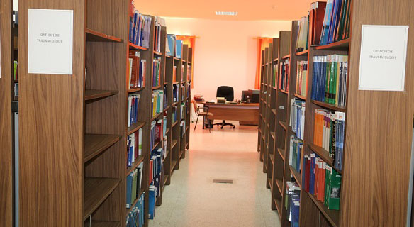
BibliothĆØque de la FMPO
CatƩgories


 Affiner la recherche Interroger des sources externes
Titre : Squire's Fundamentals of Radiology. 6th ED Type de document : texte imprimĆ© Auteurs : Robert A. Novelline, Auteur Editeur : Harvard University Press AnnĆ©e de publication : 2004 ISBN/ISSN/EAN : 978-0-674-01279-0 Langues : FranƧais (fre) CatĆ©gories : Radiologie RĆ©sumĆ© : In the past five years, the development of new imaging technologies that make possible faster and more accurate diagnoses has significantly improved the imaging of disease and injury. This new edition of Squireā€™s Fundamentals of Radiology describes and illustrates these new techniques to prepare medical students and other radiology learners to provide the most optimal and up-to-date imaging management for their patients. Not only are new diagnostic techniques outlined, such as the multidetector computed tomography diagnosis of pulmonary embolism and the diffusion-weighted magnetic-resonance imaging of stroke, but hundreds of new diagnostic images have been included to illustrate the radiological characteristics of common diseases with state-of-the-art computed radiography, ultrasound, multidetector computed tomography, and magnetic-resonance images. The text has been completely reviewed and updated to present the latest and best strategies in diagnostic imaging.
New interventional radiology procedures have been added, including vertebroplasty, a percutaneous injection treatment of painful spinal compression fractures; uterine artery embolization, a surgical alternative to hysterectomy in women with painful or bleeding uterine fibroids; and radiofrequency ablation, a percutaneous technique for treating unresectable tumors in the liver and other organs with probes that superheat and thus destroy cancer cells.
A new chapter on advances in diagnostic imaging describes many cutting-edge imaging technologies, such as three-dimensional and digital imaging, functional magnetic-resonance imaging, PETā€“CT (positron emission tomography combined with computed tomography), cardiac calcium CT scoring, multidetector gated cardiac CT, and molecular imaging.Squire's Fundamentals of Radiology. 6th ED [texte imprimĆ©] / Robert A. Novelline, Auteur . - [S.l.]Ā : Harvard University Press, 2004.
ISBN : 978-0-674-01279-0
Langues : FranƧais (fre)
CatĆ©gories : Radiologie RĆ©sumĆ© : In the past five years, the development of new imaging technologies that make possible faster and more accurate diagnoses has significantly improved the imaging of disease and injury. This new edition of Squireā€™s Fundamentals of Radiology describes and illustrates these new techniques to prepare medical students and other radiology learners to provide the most optimal and up-to-date imaging management for their patients. Not only are new diagnostic techniques outlined, such as the multidetector computed tomography diagnosis of pulmonary embolism and the diffusion-weighted magnetic-resonance imaging of stroke, but hundreds of new diagnostic images have been included to illustrate the radiological characteristics of common diseases with state-of-the-art computed radiography, ultrasound, multidetector computed tomography, and magnetic-resonance images. The text has been completely reviewed and updated to present the latest and best strategies in diagnostic imaging.
New interventional radiology procedures have been added, including vertebroplasty, a percutaneous injection treatment of painful spinal compression fractures; uterine artery embolization, a surgical alternative to hysterectomy in women with painful or bleeding uterine fibroids; and radiofrequency ablation, a percutaneous technique for treating unresectable tumors in the liver and other organs with probes that superheat and thus destroy cancer cells.
A new chapter on advances in diagnostic imaging describes many cutting-edge imaging technologies, such as three-dimensional and digital imaging, functional magnetic-resonance imaging, PETā€“CT (positron emission tomography combined with computed tomography), cardiac calcium CT scoring, multidetector gated cardiac CT, and molecular imaging.Exemplaires (1)
Code-barres Cote Support Localisation Section DisponibilitĆ© 3785 Radio 083 Livre Bibliothèque Radiologie Disponible Structure Radiologique de Base de l'OMS. Manuel d'InterprĆ©tation Radiographique pour GĆ©nĆ©ralistes / P. E. S. Palmer
Titre : Structure Radiologique de Base de l'OMS. Manuel d'InterprƩtation Radiographique pour GƩnƩralistes Type de document : texte imprimƩ Auteurs : P. E. S. Palmer, Auteur ; W. P. Cockshott, Auteur Editeur : OMS AnnƩe de publication : 1985 ISBN/ISSN/EAN : 978-92-4-254177-9 Langues : FranƧais (fre) CatƩgories : Radiologie RƩsumƩ : Preface
Acknowlegments
Chapter 1. introdction and basic concepts
Chapter 2. An invitation to think three-dimensionally
Chapter 3. How to study a chast film
Chapter 4. The lung itself
Chapter 5. The roentgen signs of lung consolidation-air space disease
Chapter 6. The diaphragm and the pleural space
Chapter 7. Mediastinal shift, atelectasis and emphysema
Chapter 8. Study of the mediastinal structures
Chapter 9. How to gather and use roentgen data
Chapter 10. The heart
Chapter 11. The abdomen: study of the plain film
Chapter 12. The abdomen: systematic examination of soft tissue zones on the plain film
Chapter 13. The abdomen: distended stomach, large and small bowel; free air and free fluid
Chapter 14. Contrast studies: principles, applications and scope
Chapter 15. Special deductions possible from excretory and secretory contrast studies
Chapter 16. Skull and bones
Structure Radiologique de Base de l'OMS. Manuel d'InterprĆ©tation Radiographique pour GĆ©nĆ©ralistes [texte imprimĆ©] / P. E. S. Palmer, Auteur ; W. P. Cockshott, Auteur . - [S.l.]Ā : OMS, 1985.
ISBN : 978-92-4-254177-9
Langues : FranƧais (fre)
CatƩgories : Radiologie RƩsumƩ : Preface
Acknowlegments
Chapter 1. introdction and basic concepts
Chapter 2. An invitation to think three-dimensionally
Chapter 3. How to study a chast film
Chapter 4. The lung itself
Chapter 5. The roentgen signs of lung consolidation-air space disease
Chapter 6. The diaphragm and the pleural space
Chapter 7. Mediastinal shift, atelectasis and emphysema
Chapter 8. Study of the mediastinal structures
Chapter 9. How to gather and use roentgen data
Chapter 10. The heart
Chapter 11. The abdomen: study of the plain film
Chapter 12. The abdomen: systematic examination of soft tissue zones on the plain film
Chapter 13. The abdomen: distended stomach, large and small bowel; free air and free fluid
Chapter 14. Contrast studies: principles, applications and scope
Chapter 15. Special deductions possible from excretory and secretory contrast studies
Chapter 16. Skull and bones
Exemplaires (2)
Code-barres Cote Support Localisation Section DisponibilitĆ© 3801 Radio 119 Livre Bibliothèque Radiologie Disponible 3802 Radio 119 Livre Bibliothèque Radiologie Disponible
Titre : TDM et IRM Cliniques : Indications et SĆ©miologie de la TomodensitomĆ©trie et de l'Imagerie par RĆ©sonance MagnĆ©tique Type de document : texte imprimĆ© Auteurs : D. Buthiau, Auteur Editeur : Frison-Roche AnnĆ©e de publication : 1992 ISBN/ISSN/EAN : 978-2-87671-160-0 Langues : FranƧais (fre) CatĆ©gories : Radiologie RĆ©sumĆ© : La TomodensitomĆ©trie et la RĆ©sonance MagnĆ©tique se placent certainement au premier rang parmi les techniques dā€™exploration diagnostique qui ont le plus bouleversĆ© nos habitudes de raisonnementā€¦
Cependant, pour ĆŖtre performants, ces procĆ©dĆ©s dā€™imagerie doivent ĆŖtre utilisĆ©s Ć bon escient, les indications correctement posĆ©es, la technique irrĆ©prochable et lā€™interprĆ©tation intelligenteā€¦
A notre Ć©poque ou le temps disponible est le bien le plus rare et le plus cher, il faut que chacun Ć©tablisse une hiĆ©rarchie dans les urgences et les besoins et une vĆ©ritable comptabilitĆ© de ses investissements intellectuelsā€¦
A ce titre, nous pensons que cet ouvrage possĆØde la qualitĆ© dā€™ĆŖtre utile au spĆ©cialiste dā€™organe ou dā€™appareil, au radiologue, au gĆ©nĆ©raliste ou Ć lā€™internisteā€¦
La rĆ©ponse ou une aide Ć la rĆ©flexion ainsi que lā€™interprĆ©tation radiologique trĆØs complĆØte et Ć©laborĆ©e pourrait ĆŖtre fournies par la lecture de ce livre dont la clartĆ© dā€™exposition est une qualitĆ© majeureā€¦
Ce livre sera, nous les prĆ©sumons, dā€™une utilisation quasi quotidienne et constituera un vĆ©ritable outil de travail et de perfectionnement.
TDM et IRM Cliniques : Indications et SĆ©miologie de la TomodensitomĆ©trie et de l'Imagerie par RĆ©sonance MagnĆ©tique [texte imprimĆ©] / D. Buthiau, Auteur . - [S.l.]Ā : Frison-Roche, 1992.
ISBN : 978-2-87671-160-0
Langues : FranƧais (fre)
CatĆ©gories : Radiologie RĆ©sumĆ© : La TomodensitomĆ©trie et la RĆ©sonance MagnĆ©tique se placent certainement au premier rang parmi les techniques dā€™exploration diagnostique qui ont le plus bouleversĆ© nos habitudes de raisonnementā€¦
Cependant, pour ĆŖtre performants, ces procĆ©dĆ©s dā€™imagerie doivent ĆŖtre utilisĆ©s Ć bon escient, les indications correctement posĆ©es, la technique irrĆ©prochable et lā€™interprĆ©tation intelligenteā€¦
A notre Ć©poque ou le temps disponible est le bien le plus rare et le plus cher, il faut que chacun Ć©tablisse une hiĆ©rarchie dans les urgences et les besoins et une vĆ©ritable comptabilitĆ© de ses investissements intellectuelsā€¦
A ce titre, nous pensons que cet ouvrage possĆØde la qualitĆ© dā€™ĆŖtre utile au spĆ©cialiste dā€™organe ou dā€™appareil, au radiologue, au gĆ©nĆ©raliste ou Ć lā€™internisteā€¦
La rĆ©ponse ou une aide Ć la rĆ©flexion ainsi que lā€™interprĆ©tation radiologique trĆØs complĆØte et Ć©laborĆ©e pourrait ĆŖtre fournies par la lecture de ce livre dont la clartĆ© dā€™exposition est une qualitĆ© majeureā€¦
Ce livre sera, nous les prĆ©sumons, dā€™une utilisation quasi quotidienne et constituera un vĆ©ritable outil de travail et de perfectionnement.
Exemplaires (1)
Code-barres Cote Support Localisation Section DisponibilitĆ© 2154 Radio 059 Livre Bibliothèque Radiologie Disponible
Titre : Teaching Atlas of Mammography. 4th Edition Type de document : texte imprimƩ Auteurs : Laszlo Tabar, Auteur ; Peter B. Dean, Auteur Editeur : Thieme AnnƩe de publication : 2012 ISBN/ISSN/EAN : 978-3-13-640804-9 Langues : FranƧais (fre) CatƩgories : Radiologie RƩsumƩ : The names Tabar and Dean are associated with high-quality mammography worldwide. In this fourth edition of the bestselling Teaching Atlas of Mammography, readers are again invited to share in the authors' experience of analyzing and evaluating mammograms. Their systematic approach not only instills a full understanding of the principles of mammography but also provides the requisite knowledge and tools to arrive at a highly accurate differential diagnosis.
Rather than starting with the diagnosis and demonstrating typical findings, the approach of this atlas is to teach the reader how to analyze the image and reach the correct diagnosis through proper evaluation of the mammographic signs. Prerequisites for the perception and evaluation of the mammographic findings are optimum technique, knowledge of anatomy, and understanding of the pathological processes leading to the mammographic appearances.
Special features :
- Revised and expanded case studies, based on 40 years of imaging experience, provide instructive long-term follow-up of patients over a period of up to 25 years
- Offers a unique comparison of imaging findings with the corresponding large thin-section and subgross thicksection (3D) histologic images to facilitate an understanding of the pathologic processes and the mammographic appearances they lead to
- Includes an abundance of coned-down compression views, microfocus magnification views, and specimen radiographs to support the analytic workup
Teaching Atlas of Mammography and the Breast Cancer book series by the same authors are essential for residents in radiology and practicing in the radiologic anatomy of the normal breast and the changes associated with benign and malignant lesions. They will help ensure that all clinicians acquire the optimal technique and knowledge of pathologic processes necessary to reach a correct diagnosis, and achieve the best long-term outcomes for their patients.Teaching Atlas of Mammography. 4th Edition [texte imprimĆ©] / Laszlo Tabar, Auteur ; Peter B. Dean, Auteur . - [S.l.]Ā : Thieme, 2012.
ISBN : 978-3-13-640804-9
Langues : FranƧais (fre)
CatƩgories : Radiologie RƩsumƩ : The names Tabar and Dean are associated with high-quality mammography worldwide. In this fourth edition of the bestselling Teaching Atlas of Mammography, readers are again invited to share in the authors' experience of analyzing and evaluating mammograms. Their systematic approach not only instills a full understanding of the principles of mammography but also provides the requisite knowledge and tools to arrive at a highly accurate differential diagnosis.
Rather than starting with the diagnosis and demonstrating typical findings, the approach of this atlas is to teach the reader how to analyze the image and reach the correct diagnosis through proper evaluation of the mammographic signs. Prerequisites for the perception and evaluation of the mammographic findings are optimum technique, knowledge of anatomy, and understanding of the pathological processes leading to the mammographic appearances.
Special features :
- Revised and expanded case studies, based on 40 years of imaging experience, provide instructive long-term follow-up of patients over a period of up to 25 years
- Offers a unique comparison of imaging findings with the corresponding large thin-section and subgross thicksection (3D) histologic images to facilitate an understanding of the pathologic processes and the mammographic appearances they lead to
- Includes an abundance of coned-down compression views, microfocus magnification views, and specimen radiographs to support the analytic workup
Teaching Atlas of Mammography and the Breast Cancer book series by the same authors are essential for residents in radiology and practicing in the radiologic anatomy of the normal breast and the changes associated with benign and malignant lesions. They will help ensure that all clinicians acquire the optimal technique and knowledge of pathologic processes necessary to reach a correct diagnosis, and achieve the best long-term outcomes for their patients.Exemplaires (1)
Code-barres Cote Support Localisation Section DisponibilitĆ© 5109 Radio 194 Livre Bibliothèque Radiologie Disponible
Titre : Techniques de Radiographie OstĆ©o-Articulaire : Savoir-Faire Type de document : texte imprimĆ© Auteurs : Maurice Rudel, Auteur ; Jean-Denis Laredo, Auteur Editeur : Sauramps MĆ©dical AnnĆ©e de publication : 2009 ISBN/ISSN/EAN : 978-2-84023-629-0 Langues : FranƧais (fre) CatĆ©gories : Radiologie RĆ©sumĆ© : La radiographie ostĆ©o-articulaire requiert des connaissances anatomiques et techniques Ć©tendues et complexes. A chaque indication correspondent des incidences spĆ©cifiques faites selon des critĆØres stricts. Ce recueil est consacrĆ© Ć toutes les incidences radiographiques ostĆ©o-articulaires hiĆ©rarchisĆ©es et dĆ©taillĆ©es en fonction de leur localisation mais aussi de leur importance. Surtout, ce recueil se veut novateur par sa prĆ©sentation trĆØs intuitive, standardisĆ©e, axĆ©e sur l'image et appuyĆ©e sur une symbolique rĆ©sumant les principaux points techniques. Le texte est synthĆ©tique, limitĆ© au strict minimum. Techniques de Radiographie OstĆ©o-Articulaire : Savoir-Faire [texte imprimĆ©] / Maurice Rudel, Auteur ; Jean-Denis Laredo, Auteur . - [S.l.]Ā : Sauramps MĆ©dical, 2009.
ISBN : 978-2-84023-629-0
Langues : FranƧais (fre)
CatĆ©gories : Radiologie RĆ©sumĆ© : La radiographie ostĆ©o-articulaire requiert des connaissances anatomiques et techniques Ć©tendues et complexes. A chaque indication correspondent des incidences spĆ©cifiques faites selon des critĆØres stricts. Ce recueil est consacrĆ© Ć toutes les incidences radiographiques ostĆ©o-articulaires hiĆ©rarchisĆ©es et dĆ©taillĆ©es en fonction de leur localisation mais aussi de leur importance. Surtout, ce recueil se veut novateur par sa prĆ©sentation trĆØs intuitive, standardisĆ©e, axĆ©e sur l'image et appuyĆ©e sur une symbolique rĆ©sumant les principaux points techniques. Le texte est synthĆ©tique, limitĆ© au strict minimum. Exemplaires (1)
Code-barres Cote Support Localisation Section DisponibilitĆ© 2338 Radio 038 Livre Bibliothèque Radiologie Disponible PermalinkPermalinkPermalink